Case study: Structure determination of pharmaceutical cocrystals
In this case study, we explored the structures of various pharmaceutical cocrystals. Due to the strong interaction of electrons with matter, 3D ED is capable of structure determination from nanometre-sized crystals and, therefore, can handle the same polycrystalline samples as PXRD whilst providing the same detail of structural information as SCXRD. For this reason, 3D ED is often employed when growing large coherent crystals for SCXRD is either unfeasible or too time-consuming.
Lacey carbon supported copper TEM grid was firstly glow- discharged for 60 s to increase the hydrophilicity of carbon film (PELCO easiGLOW, 20 mA). The pharmaceutical cocrystals were gently crushed by glass slides (VMR Microscope Slides) and delicately transferred onto pre-treated grids under the light microscope (Nikon H550L).
JEOL JEM-2100 LaB6 TEM (operated at 200kV) equipped with a fast Timepix hybrid pixel detector (512 x 512 pixels, Amsterdam Scientific Instruments) was used for 3D ED data collection. The sample crystal was continuously rotated under electron beam via the control of Instamatic software. A single tilt holder was used for the data collection under room temperature.
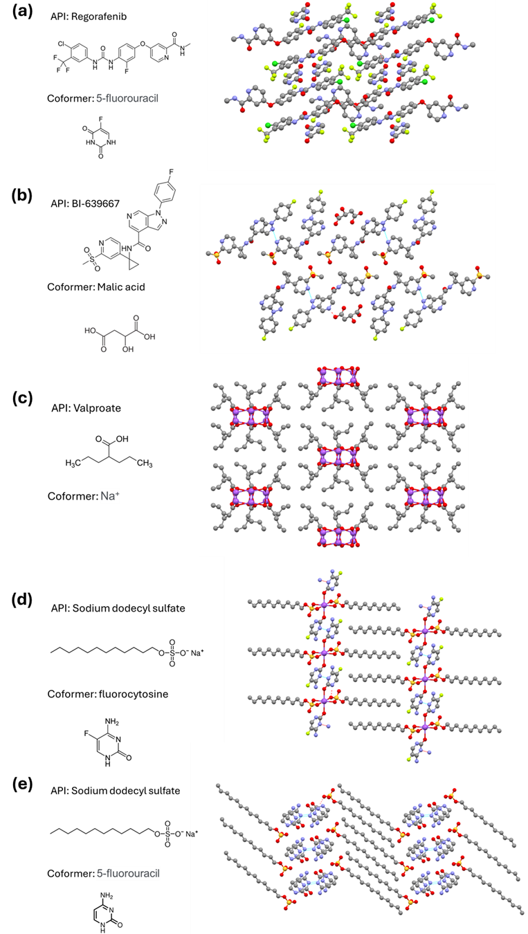
With collected cRED datasets, structures of various pharmaceutical cocrystals have been solved in this project.

The crystal packing of pharmaceutical cocrystals. (a) REG-FU; (b) : BI639667-Malic acid; (c) Sodium valproate; (d) LS fluorocytosine salt; (e) Cytosine laurate salt
