
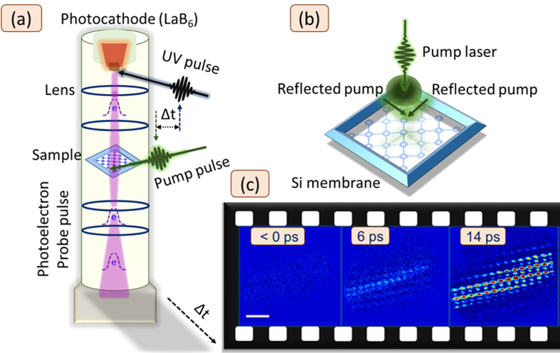
(a) Schematic illustration of the ultrafast electron microscope (UEM) used for the experimental measurements. (b) The geometry of the Si membrane [100] with slanted sides acting as mirrors. The pump laser, with a Gaussian beam profile, was carefully aligned at one of the corners of the membrane. The two perpendicular boundaries reflected the pump beam onto the surface of the membrane, forming a 2D-TG interference pattern. (c) Snapshots of the 2D-TG strain pattern resulting from ultrashort photoexcitation of the Si membrane.
Space-time visualization of structured-light-induced phonon dynamics
The ability to manipulate and explore nonequilibrium states in materials at the nanoscale is key to unlocking their functional properties. This requires high spatial and temporal resolution, as well as with possibility to transiently engineer the crystal structure, which can have a profound impact on its properties and functionality.
To achieve this, we employed ultrafast electron microscopy (UEM) combined with structured photoexcitation, specifically using two-dimensional transient grating (2D-TG) techniques. This approach allowed us to capture real-time images of strain lattices replicating the spatial profiles of 2D-TG, offering unprecedented insight into the detection and control of acoustic phonon dynamics at the nanoscale.
We observed the formation of acoustic harmonics within the acoustic cavity modes, creating a distinct frequency comb. However, under Gaussian beam excitation, this frequency comb was lifted, demonstrating the flexibility and precision of this method in controlling these dynamics. By directly visualizing and controlling these transiently engineered patterns in real space, we have introduced a powerful tool for advancing our understanding of materials and driving innovations in materials science.
The work was carried out by Amit Kumar Prasad, Gaolong Cao and Jonas Weissenrieder in KTH UEM lab.