
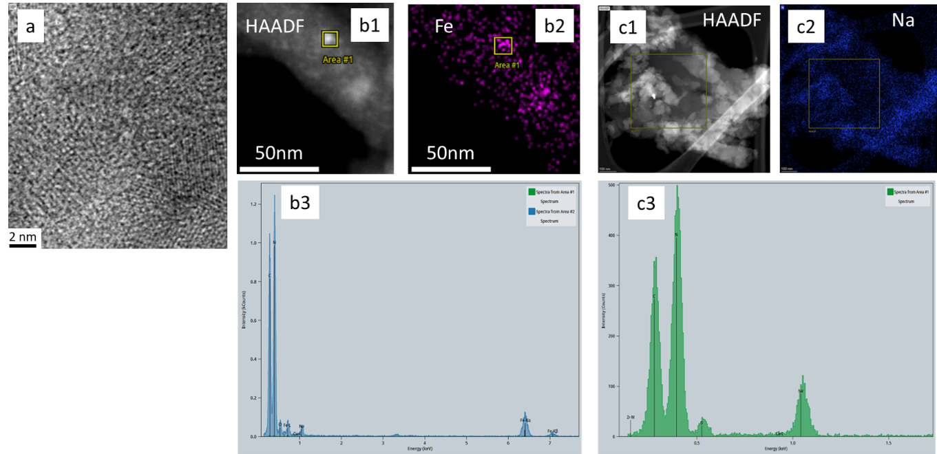
Figure: a) High resolution annular bright field image showing g-CN (002) lattice planes. The panels 1, 2, 3 are HAADF, EDS maps and EDS spectra, respectively for the Fe doped (b) and Na doped graphitic carbon nitride (c) samples.
TEM reveals the structure of modified graphitic carbon nitride photocatalysts
Transmission electron microscopy (TEM) has provided new insights into the morphology, microstructure, and elemental composition of modified graphitic carbon nitride photocatalysts developed for sustainable aviation fuel production within the EU-funded AlfaFuels project.
High-resolution TEM imaging revealed the porous and low-crystallinity nature of the materials, supporting observations obtained by X-ray diffraction (XRD). High-resolution annular bright-field imaging was also used to investigate the local crystallinity and structural features of graphitic carbon nitride.
Combined with energy-dispersive X-ray spectroscopy (EDX), TEM enabled local verification of dopant incorporation. The analysis showed distinct differences between the two investigated modifications: iron (Fe) was present as nanoparticles deposited on the carbon nitride surface, while sodium (Na) appeared to be more homogeneously distributed throughout the carbon nitride material.
These results confirm the successful synthesis and modification of the photocatalysts and provide important information for understanding how material structure, dopant distribution, and photocatalytic performance are interconnected.
The TEM investigations were performed at the Uppsala Microscopy Laboratory and contribute to the development of efficient photocatalytic materials for sustainable aviation fuel production.